
Abstract
Objectives
To estimate pooled molecular positivity for any human papillomavirus (HPV) in lesion-attributable specimens from oral potentially malignant disorders (OPMDs) and interpret findings by genotype, specimen, detection method, and viral activity.
Materials and Methods
Studies published from January 2019 to June 3, 2026 were reviewed. The meta-analysis required one independent estimate per study, lesion-directed sampling, a separable numerator and denominator, and any-HPV detection using consensus primers or broad panels. Logit-transformed proportions were pooled with a random-effects model and restricted maximum-likelihood estimation.
Results
Twenty-seven studies were included; 10 contributed 137 HPV-positive among 1,002 evaluable units. Pooled positivity was 15.7% (95% CI, 6.60–32.87; 95% prediction interval, 0.54–86.35; I² = 92.2%). Sensitivity estimates ranged from 11.4% to 21.2%. Eighteen studies yielded 357 genotype-detection events; HPV16 and HPV18 accounted for 68.3%. Twenty-five studies used lesion-associated specimens and 25 used DNA-based primary outcomes; one assessed transcription by RNA-ISH.
Conclusions
Molecular HPV detection in OPMDs is variable and method-dependent. The pooled estimate is not a uniform prevalence and does not establish oncogenic activity. Separating DNA presence, lesion attribution, and functional evidence limits unsupported causal inference.
Clinical Relevance
An isolated positive HPV DNA result is complementary. Diagnosis, dysplasia grading, and follow-up remain the basis of management; tissue localization or transcriptional testing should be reserved for a defined clinical or research question.
Keywords
- Human papillomavirus
- oral potentially malignant disorders
- oral leukoplakia
- oral lichen planus
- oral epithelial dysplasia
- meta-analysis.
Abbreviations
DNA: Deoxyribonucleic acid.
mRNA: Messenger ribonucleic acid.
CISH: Chromogenic in situ hybridization.
OSCC: Oral squamous cell carcinoma.
OPMD: Oral potentially malignant disorder.
FFPE: Formalin-fixed, paraffin-embedded tissue.
95% CI: 95% confidence interval.
PCR: Polymerase chain reaction.
PRISMA: Preferred Reporting Items for Systematic Reviews and Meta-Analyses.
qPCR: Quantitative polymerase chain reaction.
RNA-ISH: RNA in situ hybridization.
HPV: Human papillomavirus.
Introduction
Oral potentially malignant disorders (OPMDs) comprise a group of oral tissue lesions associated with a variable risk of progression to oral squamous cell carcinoma. They include oral leukoplakia, oral lichen planus, oral epithelial dysplasia, erythroplakia, oral submucous fibrosis, and other entities. Their biological behavior is heterogeneous and depends on clinical, histopathological, anatomical, behavioral, and molecular factors [1].
Human papillomavirus (HPV) has an established etiological role in a proportion of oropharyngeal carcinomas, particularly those associated with high-risk genotypes [2]. Its involvement in OPMDs, however, remains uncertain. Recent studies have reported findings ranging from no detection to high positivity in specific subgroups [3–29]. These differences may reflect the clinical entity, specimen source, diagnostic platform, and breadth of genotype coverage.
A recurring problem in the literature is the treatment of outcomes with different biological meanings as though they were equivalent. Detection of viral DNA demonstrates the presence of HPV genetic material but does not imply transcriptionally active infection, oncogene expression, or causal involvement. Likewise, an assay restricted to HPV16, HPV18, or a limited high-risk panel does not estimate positivity for any HPV [8, 11, 13–16].
Attribution of HPV to the lesion may also depend on the sampling strategy. Tissue, biopsy, or lesion-directed brushing links the finding to the affected site, whereas saliva and oral-rinse specimens may reflect HPV from other sites or transient exposure. At the molecular level, p16 is regarded as an indirect biomarker; E6/E7 mRNA detection or RNA-ISH provides evidence of transcription, whereas CISH can demonstrate tissue localization of DNA but not viral activity by itself [17–25].
The review question was: what is the proportion of molecular HPV positivity in specimens attributable to OPMD lesions, and how do genotype coverage, specimen source, and detection methods affect interpretation? The objective was to estimate this proportion and, unlike a synthesis focused solely on prevalence, preserve the reported unit of analysis, examine lesion attribution and broad viral coverage, distinguish presence, localization, transcription, and productive infection, and derive a clinical interpretive framework defining the inferences supported by each molecular result.
Materials and Methods
Study design
A systematic review with meta-analysis of proportions was conducted to evaluate HPV detection in OPMDs. Reporting followed the PRISMA 2020 statement [30].
The question was structured by population or unit of observation (patients, lesions, or specimens with OPMDs), condition (molecular HPV positivity), and context (specimens attributable to the lesion). Before synthesis, studies were classified according to specimen type, data separability, breadth of viral coverage, detection method, availability of a numerator and denominator, and risk of duplication.
The unit of analysis reported by each study—patients, lesions, specimens, or evaluable units—was retained. A single independent estimate was selected from each study for every quantitative synthesis.
Registration and protocol
This review was not registered, and no protocol was made publicly available.
Eligibility criteria
Original human studies published in English or Spanish from January 2019 through June 3, 2026 that assessed HPV in OPMDs were eligible. The time restriction was applied to synthesize contemporary evidence. Oral leukoplakia, oral lichen planus, oral epithelial dysplasia, erythroplakia, oral submucous fibrosis, oral lichenoid lesions, proliferative verrucous leukoplakia, and other entities classified by the authors as OPMDs were considered.
The descriptive synthesis accepted data on HPV presence, genotypes, methods, p16, and transcriptional or tissue-level evidence. The primary meta-analysis required a specimen attributable to the lesion, separable OPMD data, identifiable numerator and denominator, general HPV detection using consensus primers or a broad multigenotype panel, and one independent estimate. Assays directed only at a predefined selection of genotypes were not considered equivalent to detection of any HPV.
Studies were excluded from the primary meta-analysis if they relied exclusively on saliva, oral rinse, or other specimens not attributable to the lesion; used p16 without molecular HPV detection; assessed only one or several preselected genotypes without estimating any-HPV positivity; did not permit separation of OPMDs from carcinoma, controls, or other lesions; provided an insufficient diagnostic or molecular definition to verify the outcome; lacked an extractable numerator or denominator; contained internal inconsistencies that precluded identification of a unique estimate; or potentially duplicated another cohort without an independent estimate that could be selected.
Informative studies that did not meet the criteria for the primary analysis were retained for narrative synthesis or non-poolable qualitative synthesis according to the type and comparability of their data. Their quantitative results were described without incorporation into the estimate of any-HPV positivity.
Information sources and search strategy
The search was conducted in PubMed/MEDLINE, Google Scholar, OpenAlex, and the Cochrane Library and was last updated on June 3, 2026. HPV terms were combined with OPMDs and their principal clinical diagnoses.
The base search string was: ("human papillomavirus" OR HPV OR "HPV DNA" OR "HPV genotype") AND ("oral potentially malignant disorders" OR "oral premalignant lesions" OR "oral leukoplakia" OR "oral lichen planus" OR "oral epithelial dysplasia" OR "oral submucous fibrosis" OR erythroplakia OR "oral mucosa" OR "oral cavity"). Reference lists of included studies and relevant reviews were also examined. The reproducible strategy for each source is provided in Online Resource 1.
Study selection
Records were integrated into a single matrix. Duplicates were identified using title, authors, year, journal, DOI, or another available identifier.
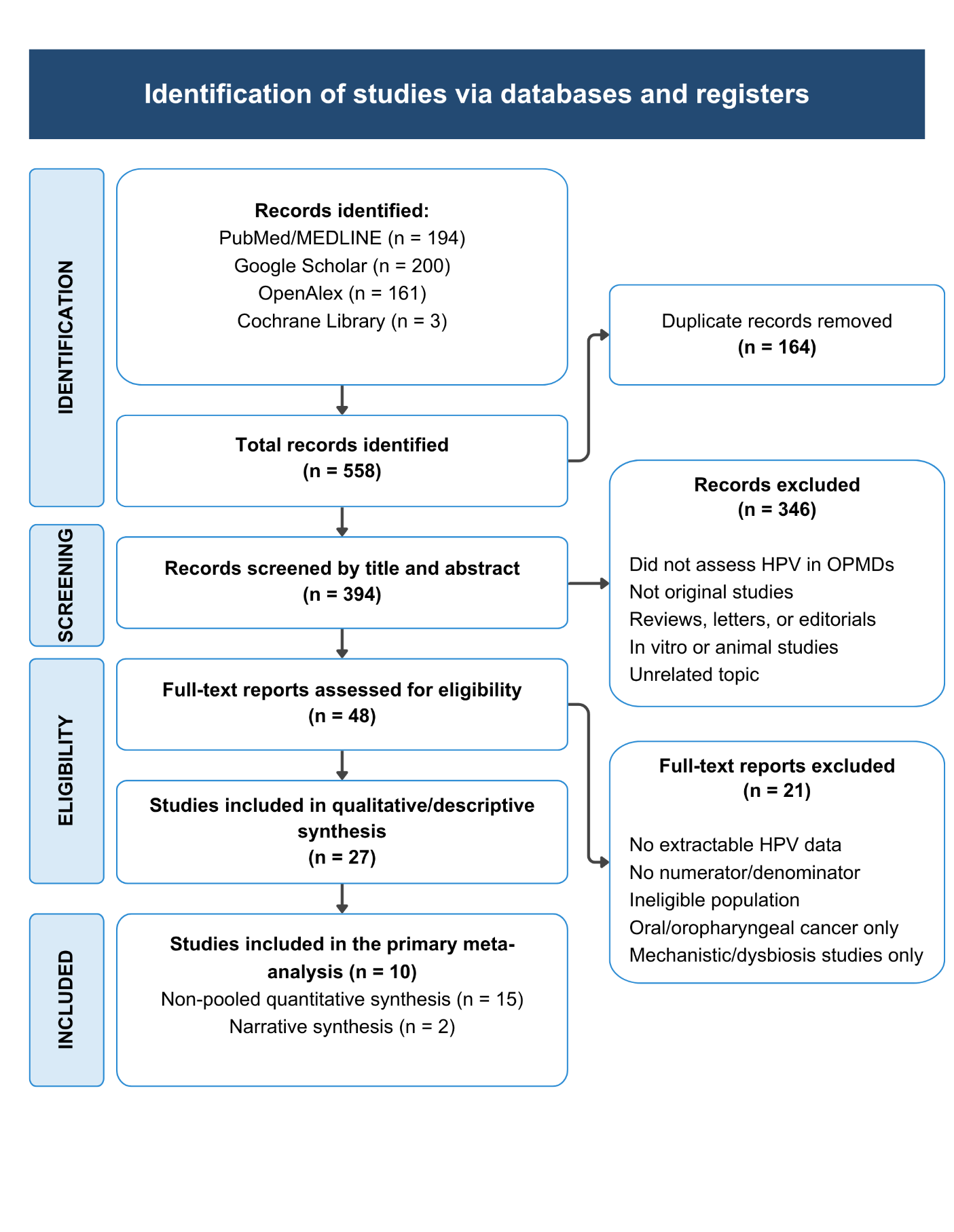
Selection was performed in two stages: title and abstract screening followed by full-text review. During full-text review, an analytical role was assigned to each study. The retained screening log verified 21 full-text exclusions and five general exclusion categories, but the number within each category could not be reconstructed reliably; frequencies were therefore not imputed. The complete process is shown in Fig. 1.
One reviewer performed the initial screening, and a second reviewer verified the decisions. Disagreements were resolved by joint full-text review and consensus.
Data extraction
A structured matrix was used to record author, year, country, design, OPMD diagnosis, diagnostic criteria, sample size, unit of analysis, specimen, method, viral panel, evaluable units, positive and negative counts, genotypes, indirect biomarkers, and transcriptional or tissue-level evidence.
When a study reported multiple methods, lesions, or specimens from the same participants, a single estimate was selected. Priority was given to lesion attribution, separability of OPMD data, the complete denominator, direct molecular detection, and avoidance of double counting.
Genotypes were retained as reported. Combined categories such as HPV6/11 or HPV16/18 were not separated. Counts were treated as detection events rather than unique patients unless the study demonstrated that no overlap occurred.
One reviewer performed the initial extraction, and a second reviewer verified the matrix. Disagreements were resolved by consulting the original article.
Outcomes
The primary outcome was the proportion of evaluable units with molecular detection of any HPV in a specimen attributable to the OPMD:
HPV-positive units / total evaluable units.
Secondary outcomes were reported genotypes, specimen types, detection platforms, breadth of viral coverage, sensitivity analyses, and narrative evidence on p16, tissue localization, or viral activity.
Viral DNA was interpreted as the presence of genetic material; p16 as an indirect biomarker; E6/E7 mRNA or RNA-ISH as transcriptional evidence; DNA CISH as tissue localization; and E4/L1, when applicable, as markers of productive infection. These outcomes were not pooled together. Based on this hierarchy and the clinical and histopathological management of OPMDs [1], a framework for clinical interpretation was developed. The framework is not a validated diagnostic rule and does not assign transformation risk; it organizes the scope and limitations of each molecular result.
Analytical classification
Studies were organized according to their primary analytical use: meta-analysis, narrative synthesis, or non-poolable qualitative synthesis. The meta-analysis included estimates of any HPV obtained from specimens attributable to the lesion, with complete and independent data and a broad-coverage method. The narrative synthesis summarized biomarkers and functional evidence; the qualitative synthesis retained relevant findings whose panel, specimen, diagnostic definition, internal consistency, or dependence between cohorts precluded incorporation into the primary estimate.
Critical appraisal and risk of bias
Critical appraisal was performed using the Joanna Briggs Institute checklist for prevalence studies [31]. Nine domains were assessed: appropriateness of the sampling frame, participant selection, sample size, description of participants and setting, sample coverage, validity of the identification method, consistency of measurement, statistical analysis, and response rate or management of an inadequate response. Each domain was judged as “yes,” “no,” “unclear,” or “not applicable.”
The initial appraisal was performed by one reviewer and the matrix was verified by a second reviewer; disagreements were resolved by consulting the primary article and reaching consensus. Judgments were presented by domain. No overall score was calculated, and the JBI checklist was not converted into overall categories using unvalidated thresholds. Appraisal was not used as an automatic exclusion criterion.
Statistical synthesis
For each study, the evaluable total and the HPV-positive and HPV-negative counts were extracted. Study-specific positivity was calculated as events divided by total.
The meta-analysis was implemented in R using explicit inverse-variance code applied to logit-transformed proportions. Between-study variance (τ²) was estimated by restricted maximum likelihood (REML), the primary 95% CI was obtained using a normal approximation, and estimates were back-transformed to proportions. To assess robustness given the small number of studies and substantial heterogeneity, the 95% CI was recalculated using the Hartung–Knapp–Sidik–Jonkman adjustment with a t distribution and k−1 degrees of freedom.
Study-specific 95% CIs were calculated using the Clopper–Pearson method. When a scenario included a study with zero events, 0.5 was added to the event and non-event cells of all studies in that scenario (method.incr = "all"). The analysis excluding the zero-event study assessed the influence of this decision.
Heterogeneity was quantified using I², τ² on the logit scale, and Cochran’s Q. A 95% CI for the pooled estimate and a 95% prediction interval were calculated using a t distribution with k−1 degrees of freedom. The sum of events divided by the sum of units was reported only as descriptive crude positivity and not as the meta-analytic estimate.
Forest plots displayed study-specific and pooled estimates. Given the number of studies, heterogeneity, and the mathematical dependence between a proportion and its standard error, the funnel plot was treated only as an exploration of small-study effects and not as a test of publication bias.
Sensitivity analyses
In addition to the primary scenario, three data-composition sensitivity analyses were performed (tissue or biopsy only, exclusion of the zero-event study, and exclusion of the largest study), together with an inferential check using the Hartung–Knapp–Sidik–Jonkman adjustment. Meta-regression and pooled estimates by diagnosis were not performed because too few studies were available per covariate and several series included overlapping clinical categories.
Small-study effects and missing results
A funnel plot was inspected for the ten studies in the primary analysis. No formal asymmetry tests were applied, and any observed asymmetry was not attributed to publication bias.
Certainty of evidence
No formal certainty-of-evidence assessment was performed.
Results
Study selection
A total of 558 records were identified: 194 (34.8%) in PubMed/MEDLINE, 200 (35.8%) in Google Scholar, 161 (28.9%) in OpenAlex, and 3 (0.5%) in the Cochrane Library. After 164 duplicates (29.4%) were removed, 394 records were screened; 346/394 (87.8%) were excluded by title or abstract. Of 48 full-text reports, 21 (43.8%) were excluded and 27 (56.3%) entered the descriptive synthesis.
Twenty-five of 27 studies (92.6%) reported a numerator and denominator for at least one positivity outcome. Ten (37.0%) met the criteria for the primary meta-analysis, 15 (55.6%) contributed non-poolable quantitative findings, and 2 (7.4%) were retained for narrative synthesis.
General characteristics of the included studies
The characteristics of the 27 studies are presented in Table 1. Because units of analysis differed and some cohorts were known to overlap, denominators were not summed as though they represented unique participants.
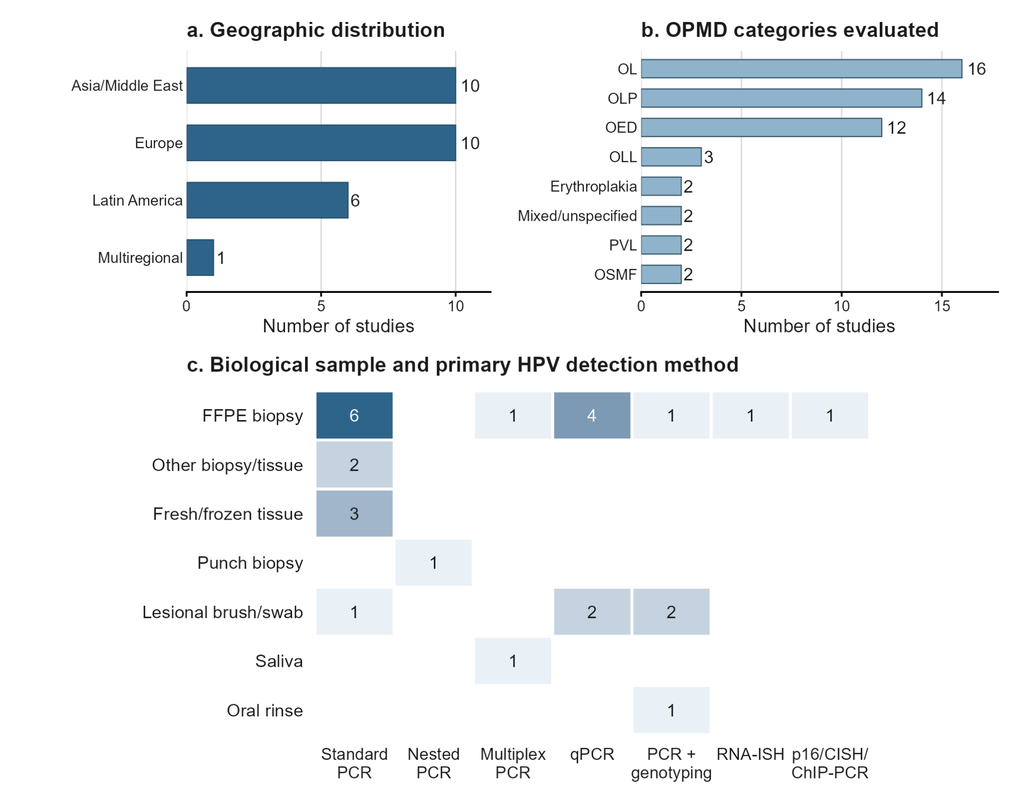
By region, 10/27 studies (37.0%) were conducted in Europe, 10/27 (37.0%) in Asia or the Middle East, 6/27 (22.2%) in Latin America, and 1/27 (3.7%) was multinational. Twelve (44.4%) were cross-sectional, 8 (29.6%) retrospective or archival, 5 (18.5%) prospective, and 2 (7.4%) case-control studies.
Clinical categories were not mutually exclusive. Sixteen of 27 studies assessed oral leukoplakia (59.3%), 14/27 oral lichen planus (51.9%), and 12/27 oral epithelial dysplasia (44.4%); 3/27 included oral lichenoid lesions (11.1%), and 2/27 each (7.4%) included erythroplakia, mixed or unspecified categories, oral submucous fibrosis, and proliferative verrucous leukoplakia.
Twenty of 27 studies (74.1%) used tissue or biopsy as the primary specimen: 14 FFPE specimens (51.9%), 3 fresh or frozen specimens (11.1%), 2 other biopsy or tissue specimens (7.4%), and 1 punch biopsy (3.7%). Five studies (18.5%) used lesion-directed brushing, swabbing, or exfoliated cells; 1 (3.7%) analyzed saliva and 1 (3.7%) oral rinse.
The primary viral outcome was DNA based in 25/27 studies (92.6%); these studies used conventional, nested, or multiplex PCR, qPCR, and genotyping platforms. Hendawi et al. used RNA-ISH (1/27; 3.7%), and Parchami et al. used a staged algorithm combining p16, CISH, and ChIP-PCR (1/27; 3.7%). The descriptive profile by region, OPMD category, specimen, and method is shown in Fig. 2.
Critical appraisal and risk of bias
The JBI domains with the greatest uncertainty were participant selection (22/27 “unclear”; 81.5%), adequacy of sample size (11/27 “no”; 40.7%, and 5/27 “unclear”; 18.5%), and response rate or its management (17/27 “unclear”; 63.0%). In contrast, 26/27 studies (96.3%) satisfied each of the domains concerning description of the setting, validity of the method, consistency of measurement, and appropriateness of analysis. Of 243 domain-level judgments, 178 (73.3%) were “yes,” 53 (21.8%) “unclear,” and 12 (4.9%) “no.” The post-appraisal decision was “include” for 26/27 studies and “seek further information” for 1/27; this decision is not an overall risk-of-bias category. Complete judgments are presented in Online Resource 2.
Analytical classification
Ten studies (37.0%) contributed to the primary meta-analysis, representing 1,002 evaluable units: 137 positive and 865 negative. Fifteen additional studies (55.6%) were retained as non-poolable quantitative evidence and 2 (7.4%) as narrative evidence (Table 2).
The 17 studies not pooled were excluded from the estimate because of a restricted panel, a non-lesional specimen, lack of a separable global estimate, internal inconsistency, or dependence between cohorts; their findings remained visible without being pooled.
Primary meta-analysis of HPV positivity
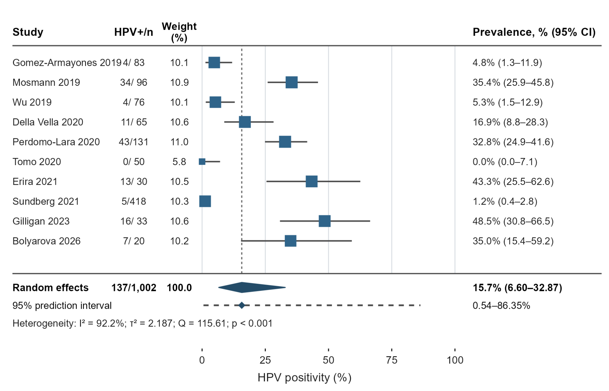
The ten studies comprised 1,002 evaluable units, including 137 positive and 865 negative units. Study-specific positivity ranged from 0.0% to 48.5%; cumulative crude positivity was 137/1,002 (13.7%) and is reported for descriptive purposes only (Table 3).
The pooled proportion positive for any HPV was 15.7% (95% CI 6.60–32.87). The 95% prediction interval was 0.54–86.35%, spanning 85.81 percentage points. Heterogeneity was very high (I² = 92.2%; τ² = 2.187 on the logit scale; Q = 115.61; p < 0.001). Figure 3 shows the study-specific and pooled estimates.
Sensitivity analyses
Table 4 summarizes three data-based sensitivity analyses and an inferential check using the Hartung–Knapp adjustment. Relative to the primary estimate of 15.7%, alternative scenarios ranged from −4.3 to +5.5 percentage points and retained high or very high heterogeneity (I² = 85.5–92.7%). These differences are not formal subgroup comparisons.
The tissue- or biopsy-only analysis included 8 studies and 87 positive units among 873 evaluable units (10.0% crude positivity). Pooled positivity was 11.4% (95% CI 4.07–27.91), 4.3 percentage points below the primary estimate; the 95% prediction interval was 0.29–84.98%, and I² was 92.7% (τ² = 2.248; Q = 95.84; Online Resource 3).
After exclusion of the zero-event study, 9 studies remained, with 137 positive units among 952 evaluable units (14.4% crude positivity). Pooled positivity was 17.7% (95% CI 7.48–36.29), 2.0 percentage points above the primary estimate; the 95% prediction interval was 0.63–87.87%, and I² was 92.5% (τ² = 2.082; Q = 106.71; Online Resource 4).
After exclusion of the largest study, 9 studies remained, with 132 positive units among 584 evaluable units (22.6% crude positivity). Pooled positivity was 21.2% (95% CI 10.86–37.16), 5.5 percentage points above the primary estimate; the 95% prediction interval was 1.73–80.36%, and I² was 85.5% (τ² = 1.233; Q = 55.15; Online Resource 5).
The Hartung–Knapp adjustment retained the point estimate of 15.7% but widened the 95% CI from 6.60–32.87% to 5.56–37.01%. The substantive conclusion was unchanged: imprecision was considerable and compatible with markedly different positivity values.
Small-study effects
The funnel plot included the 10 studies in the primary analysis (Online Resource 6). Given k = 10, I² = 92.2%, and the mathematical dependence between a proportion and its standard error, formal asymmetry tests were not applied and visual inspection was not interpreted as evidence of publication bias.
Genotype distribution
Eighteen of 27 studies (66.7%) contributed usable genotype data: 357 detection events in 30 viral or non-separable categories (Online Resource 7). Of these, 316 (88.5%) came from specimens attributable to the lesion and 41 (11.5%) from saliva; the two strata were not considered directly comparable.
Because studies used different oncogenic-risk classifications for some genotypes, the 357 events were not redistributed into aggregated risk categories. The synthesis retains the original viral labels and non-separable categories.
HPV16 accounted for 157/357 events (44.0%) and HPV18 for 87/357 (24.4%); together they represented 244/357 events (68.3%). They were followed by HPV6 (22; 6.2%), HPV11 (14; 3.9%), HPV49 (9; 2.5%), and HPV33 (8; 2.2%). These proportions describe reported detection events, not genotype-specific prevalence.
Genotype counts do not correspond to unique patients: several studies allowed codetection and others assessed restricted panels. Therefore, neither pooled genotype-specific prevalence nor the proportion of single and multiple infections was estimated. Online Resource 8 retains the study, specimen, method, and non-separable categories.
Detection methods and specimen type
Twenty-five of 27 studies (92.6%) used specimens attributable to the lesion: 20 (74.1%) used tissue or biopsy and 5 (18.5%) used lesion-directed brushing, swabbing, or exfoliated cells. Two studies (7.4%) used non-lesional specimens: one saliva sample and one oral-rinse sample.
Primary detection was DNA based in 25/27 studies (92.6%). Conventional, nested, and multiplex PCR, qPCR, and genotyping platforms with different breadths of coverage were used; equivalent diagnostic sensitivity was therefore not assumed.
Hendawi et al. used RNA-ISH as transcriptional evidence, whereas Parchami et al. used a staged algorithm combining p16, CISH, and ChIP-PCR. These outcomes were kept separate from general DNA positivity.
Biomarkers and functional evidence
Ten of 27 studies (37.0%) contributed complementary biomarkers or viral/epigenetic evidence (Online Resource 9). Nine reported p16 using non-uniform thresholds. Only Hendawi et al. quantified transcription using RNA-ISH (6/33; 18.2%); Parchami et al. reported CISH in 5/54 cases (9.3%) and ChIP-PCR in 3/9 selected cases, without extrapolating that subset.
Indirect biomarkers, tissue localization, and transcriptional evidence were not treated as equivalent and were not incorporated into the primary estimate unless the study also reported an eligible independent molecular measurement.
Discussion
Principal findings and contribution of this review
The central finding is not merely a pooled positivity of 15.7%, but its marked instability across settings. Study-specific estimates ranged from 0.0% to 48.5%, the 95% CI was 6.60–32.87%, and the 95% prediction interval was 0.54–86.35%. Together with I² = 92.2%, these findings indicate that a new clinical setting may observe positivity that differs substantially from the meta-analytic mean and that 15.7% should not be communicated as a universal prevalence for OPMDs.
Only 10/27 studies (37.0%) contributed estimates compatible with any-HPV positivity in specimens attributable to the lesion. The remaining 17 studies were not discarded: they were retained to show how restricted panels, non-lesional specimens, dependent cohorts, non-separable denominators, and indirect biomarkers alter the meaning of a result. This classification is itself a methodological finding because it avoids creating apparent precision by combining biologically different outcomes.
The principal contribution of this review is therefore interpretive harmonization. A single independent estimate was selected per study; patients, lesions, specimens, and genotype-detection events were distinguished; and evidence was ordered from DNA presence to tissue localization, transcription, and productive infection. This approach shifts the question from “How much HPV is present?” to “What did each study actually measure, and what inference does that measurement support?”
Oral leukoplakia (59.3%), oral lichen planus (51.9%), and oral epithelial dysplasia (44.4%) were the most frequently represented entities, but categories overlapped and several series combined diagnoses. Consequently, the estimate describes the analyzable set rather than a specific lesion. The lack of sufficient independent estimates by diagnosis precludes application of the 15.7% estimate to any particular clinical entity.
Comparison with previous evidence
The meta-analysis by de la Cour et al. included 52 studies and 2,677 cases and estimated HPV DNA prevalence at 22.5% (95% CI 16.6–29.0; I² = 93%) [32]. The contemporary estimate in the present review was 15.7%, 6.8 percentage points lower. However, overlap between the confidence intervals and differences in eligibility criteria preclude interpretation as a temporal decline. The difference may reflect the 2019–2026 period, the requirement for lesion attribution, the exclusion of restricted panels as estimators of any HPV, and selection of a single independent estimate per study.
The nearly identical heterogeneity (92.2% vs 93%) is more informative than the difference between pooled estimates: the introduction of more recent molecular platforms has not resolved the lack of standardization in diagnosis, sampling, processing, assay coverage, and unit of analysis. Technology has increased detection capacity but not necessarily clinical comparability across studies.
HPV16 represented 48.2% of HPV-positive cases in the previous review and 44.0% of genotype-detection events in the present synthesis [32]. This numerical similarity does not imply equivalence. The former denominator consisted of positive cases; the latter consisted of detection events with possible codetections and different panels. Preserving this distinction prevents presentation of the event composition as individual genotype prevalence.
Sources of heterogeneity and generalizability
Heterogeneity probably reflects the interaction of clinical and technical differences. Studies included entities with different biological risks, populations from diverse regions, and diagnostic criteria that were not always equivalent. Denominators also represented patients, lesions, specimens, or evaluable units. A participant may contribute more than one lesion, and a genotype codetection is not a new patient; retaining the original unit reduced double counting but did not eliminate this structural variation.
Specimen source affected attribution of the finding. Twenty-five studies used tissue or lesion-directed sampling, whereas two analyzed saliva or oral rinse. Restricting the meta-analysis to tissue or biopsy reduced the estimate to 11.4%, although the persistently wide prediction interval precludes attribution of the difference solely to specimen type. In the 14 studies using FFPE material, fixation, storage, DNA fragmentation, and the length of the amplified region may have influenced sensitivity, but these factors were not reported uniformly [33].
Assay breadth was another major source of variation. Conventional, nested, and multiplex PCR, qPCR, and genotyping platforms do not necessarily share limits of detection or coverage. An assay directed only at HPV16/18 may answer a specific etiological question but does not estimate positivity for any HPV. Insufficient data precluded defensible meta-regression by region, diagnosis, specimen, or platform; sources of heterogeneity are therefore interpreted as hypotheses supported by the pattern across studies rather than quantified effects.
Biological interpretation of molecular outcomes
Detection of DNA confirms the presence of HPV genetic material but does not demonstrate viral integration, oncogene transcription, or causal involvement. Positivity may represent persistent infection, incidental presence, transient exposure, or surface contamination. This distinction is particularly important in the oral cavity, where the model of HPV-associated oropharyngeal carcinoma should not be extrapolated automatically [2].
Outcomes can be ordered according to the inference they support. CISH provides tissue localization of DNA; E6/E7 mRNA or RNA-ISH demonstrates transcription; and E4/L1 supports productive infection. Even these levels do not establish by themselves that the virus drives malignant progression; temporal association, persistence, molecular plausibility, and longitudinal outcomes are required. In the available evidence, only 1/27 studies used RNA-ISH as the primary outcome and detected transcription in 6/33 lesions [17].
p16 should not be used as an isolated surrogate for HPV in OPMDs. Tomo et al. observed high p16 expression in 20/50 leukoplakias with detectable HPV DNA in 0/50 [19], whereas Hendawi et al. reported p16 in 11/33 lesions and RNA-ISH positivity in 6/33 [17]. This discordance, together with non-uniform immunohistochemical thresholds, confirms that p16, DNA, and transcription address different questions.
Framework for clinical interpretation
Figure 4 translates these findings into an interpretive clinical algorithm. The starting point remains unchanged: clinical assessment, biopsy when indicated, histopathological diagnosis, dysplasia grading, and follow-up determine OPMD management [1]. This review found insufficient evidence to recommend routine HPV screening or to modify treatment or surveillance on the basis of an isolated molecular result.
When a specific clinical or scientific question exists, testing should use a lesion-directed specimen and an assay with documented target region, coverage, limit of detection, and controls. A negative result indicates only that DNA was not detected with that specimen and assay; a positive result confirms viral presence, not causality. Saliva or oral-rinse specimens and restricted panels must be interpreted within their limits of attribution and coverage.
Localization or activity assays may refine biological interpretation but are not validated prognostic markers. HPV status should therefore be recorded as a complementary molecular phenotype rather than as a clinical category capable of displacing histopathology. The proposed framework is intended to prevent overinterpretation; it is neither a clinical practice guideline nor a validated decision tool.
Strengths and limitations
Strengths include pooling criteria defined by lesion attribution, broad viral coverage, separability, and independence; selection of one estimate per study; preservation of non-separable genotype categories; exact study-specific confidence intervals; a prediction interval; three sensitivity analyses; a Hartung–Knapp check; and domain-level JBI appraisal without conversion to an unvalidated overall score. These decisions made uncertainty visible rather than concealing it behind a single summary estimate.
Limitations of the evidence were quantifiable: 22/27 studies did not clearly describe selection, 16/27 had an inadequate or uncertain sample size, and 17/27 did not adequately report response rate or its management. Only 10 studies could be pooled and heterogeneity was very high; robust diagnosis-specific estimates and meta-regression were therefore not performed. The pooled estimate does not represent population prevalence and retains different units of analysis across studies.
The search was restricted to 2019–2026, English or Spanish, and four sources; it did not include Embase, Scopus, or Web of Science, and only the first 200 Google Scholar results ranked by relevance were screened. Screening and extraction were verified but were not performed completely independently by two reviewers. These decisions may have reduced comprehensiveness and reproducibility.
In addition, 25/27 studies primarily assessed DNA, functional evidence was sparse, and no formal certainty-of-evidence assessment was applied. Panels considered broad were not identical. Finally, the clinical framework was derived from the synthesis and the biological hierarchy of outcomes but has not been prospectively validated as a decision tool.
Research implications
Future studies should adopt a minimum reporting set comprising the clinical and histopathological definition of the lesion, anatomical site, unit of analysis, specimen source, pre-analytical conditions, target region and amplicon length, limit of detection, quality controls, complete list of genotypes covered by the panel, and codetections by participant and lesion.
Longitudinal designs should link lesion-associated detection with persistence and transformation, integrate DNA with E6/E7 mRNA or RNA-ISH, tissue localization, and markers of productive infection, and determine whether this information adds prognostic value beyond clinical assessment and histopathology. Without temporal outcomes, molecular plausibility cannot be converted into clinical causality.
Standardization of units and assays would enable comparisons across regions, diagnoses, and platforms, reduce heterogeneity, and determine whether a reproducible subgroup of OPMDs exists in which HPV has additional biological or clinical value.
Conclusions
Across 10 studies and 1,002 evaluable units, pooled positivity for any HPV was 15.7%; however, the 95% prediction interval of 0.54–86.35% and I² of 92.2% preclude interpretation as a uniform prevalence or application to a specific OPMD category.
HPV16 and HPV18 accounted for 68.3% of genotype-detection events, although these counts depend on panel composition, allow codetection, and do not represent unique patients or individual genotype prevalence.
Contemporary evidence shows that DNA detection does not establish active infection, causality, or transformation risk. Clinical management should remain grounded in clinical assessment and histopathology; HPV testing provides only complementary information within a defined question.
Well-characterized lesion-associated specimens, sufficiently broad assays, independent units, functional markers, and longitudinal follow-up are required to determine whether HPV provides additional biological or clinical value in OPMDs.
Figures
![Fig. 4 Proposed framework for clinical interpretation of HPV testing in OPMDs. The algorithm organizes but does not replace clinical and histopathological assessment. DNA identifies viral material; p16 is indirect, CISH localizes DNA, RNA-ISH or E6/E7 mRNA demonstrates transcription, and E4/L1 supports productive infection. No single level demonstrates causality or justifies treatment changes. Developed from the outcome hierarchy in this review and the clinical and histopathological management of OPMDs [1]](/volumenes/biohorizons/1/assets/figure-4.png)
Table 1. General characteristics of the included studies
| Authors and year | Country | Design | OPMD | Specimen | Detection method | Reported viral result | n | HPV+ |
|---|---|---|---|---|---|---|---|---|
| Gomez-Armayones et al., 2019 | Spain | R/CS | OLP; OED | FFPE biopsy | SPF10 PCR-DEIA/LiPA25 | HPV DNA + genotyping | 83 | 4 |
| Mosmann et al., 2019 | Argentina | CS | Mixed OPMDs, NS | Lesion-directed swab | PCR MY09/MY11 + RFLP | HPV DNA + genotyping | 96 | 34 |
| Sundberg et al., 2019 | Sweden | P/CS | OL | FFPE biopsy | Real-time TaqMan PCR | HPV DNA; 13-type panel | 74 | 0 |
| Wu et al., 2019 | China | R | OL | FFPE biopsy | Reverse dot blot + HPV16 PCR | HPV DNA; 23 types (only HPV16 detected) | 76 | 4 |
| Della Vella et al., 2020 | Italy | P | OL | Lesional microbiopsy | Anyplex II HPV28 (real-time PCR) | HPV DNA; 28-type panel | 65 | 11 |
| Farhadi et al., 2020 | Iran | R/CS | OLP | FFPE biopsy | Multiplex/genotype-specific PCR | HPV16/18/33 DNA | 32 | 8 |
| Hendawi et al., 2020 | United Kingdom | R/CS | OED | FFPE biopsy | RNAscope RNA-ISH for HPV16/18 E6/E7 | HPV16/18 E6/E7 RNA | 33 | 6 |
| Perdomo-Lara et al., 2020 | Colombia | CS | OED | FFPE biopsy | qPCR GP5+/GP6+ + Luminex xMAP | HPV DNA; 24-type panel | 131 | 43 |
| Tomo et al., 2020 | Brazil | CS | OL | Fresh/frozen tissue | PCR PGMY09/11 + Linear Array | HPV DNA; broad panel | 50 | 0 |
| Buenahora et al., 2021 | Colombia | P/CS | OED in OL/OLP/erythroplakia | FFPE biopsy | qPCR/Luminex; p16 IHC; RRBSᵃ | HPV16/p16/methylation; inconsistent countᵃ | 36–37ᵃ | NRᵃ |
| Erira et al., 2021 | Colombia | CS | OL with OED | FFPE biopsy | Conventional GP5+/GP6+ PCR | HPV DNA; HPV16 confirmation | 30 | 13 |
| Ghosh et al., 2021 | Nepal | R/CS | OED; OLP; OSMF | FFPE biopsy | Conventional HPV16/18 PCR | HPV16/18 DNA | 150 | 2 |
| Kaewmaneenuan et al., 2021 | Thailand | R/CS | OL; OLP | FFPE biopsy | Conventional HPV16/18 E6 PCR | HPV16/18 DNA | 160 | 31 |
| Runow Stark et al., 2021 | Sweden | P/CS | Mixed OPMD/non-OPMD lesionsᵉ | Fresh biopsyᵉ | TaqMan PCR (12 HR types + HPV6/11)ᵉ | HR- and LR-HPV DNAᵉ | 46ᵉ | 0ᵉ |
| Sivakumar et al., 2021 | India | P/CS | OL | Lesion-directed cytobrush | HPV16 E6-specific PCR | HPV16 DNA | 25 | 5 |
| Sundberg et al., 2021 | Sweden/Brazil/Romania | R | OL | FFPE biopsy | Real-time TaqMan PCR | HPV DNA; 14-type panel | 418 | 5 |
| Rathore et al., 2022 | India | R/CS | Mixed OPMDs, NS | FFPE biopsy | HPV DNA PCR | HPV DNA; genotype NS | 30 | 0 |
| Vijayan et al., 2022 | India | CS | OLP | Biopsy/fresh tissue | Conventional HPV16/18 E6 PCR | HPV16/18 DNA | 250 | 103 |
| Fantozzi et al., 2024 | Italy | CS | Mixed OPMDs | Lesion-directed brushing | Real-time/genotype-specific PCR | HPV DNA + complementary p16 | 59 | 12 |
| Gilligan et al., 2023 | Argentina | CS | OL; PVL | Lesion-directed brushing | PCR MY09/11 + GP5+/GP6+; RFLP | HPV DNA + genotyping | 33 | 16 |
| Mohammadi et al., 2023 | Iran | CC | OLP | FFPE biopsy | Conventional HPV16/18 PCR | HPV16/18 DNA | 25 | 14 |
| Petrović et al., 2023 | Serbia | CS | OL; OLP | Lesion-directed swab | qPCR for 12 HR genotypes | HR-HPV DNA | 30 | 2 |
| Parchami et al., 2024 | Iran | CS | OED | FFPE biopsy | p16 IHC + CISH + ChIP-PCRᶜ | CISH 5/54; ChIP-PCR 3/9 selected casesᶜ | 54 | NRᶜ |
| Buttà et al., 2025 | Italy | CS | OL; OLP; erythroplakia | Oral rinseᵈ | INNO-LiPA; PCR/Sangerᵈ | HPV DNA in oral-rinse specimensᵈ | 17ᵈ | 7ᵈ |
| Duś-Ilnicka et al., 2025 | Poland | CS | OLP | Salivaᵇ | E7-MPG PCR + Luminexᵇ | Salivary genotypes; no overall participant-level resultᵇ | 31ᵇ | NRᵇ |
| Bolyarova et al., 2026 | Bulgaria | CS | Mixed OPMDs | Punch biopsy | Nested PCR + hybridization/chip | HPV DNA + genotyping | 20 | 7 |
| Elsahn et al., 2026 | UAE | CC | OL; OLP; OSMF | Fresh tissue biopsy | HPV16 E6-specific PCR | HPV16 DNA | 60 | 11 |
Abbreviations: CC, case-control; CISH, chromogenic in situ hybridization; CS, cross-sectional; FFPE, formalin-fixed, paraffin-embedded tissue; HPV, human papillomavirus; HR, high risk; IHC, immunohistochemistry; LR, low risk; NR, not reportable; NS, not specified; OED, oral epithelial dysplasia; OL, oral leukoplakia; OLP, oral lichen planus; OPMD, oral potentially malignant disorder; OSMF, oral submucous fibrosis; P, prospective; PVL, proliferative verrucous leukoplakia; R, retrospective; UAE, United Arab Emirates. Note: n evaluated is the denominator for the viral measurement shown and not necessarily the total sample size of the article. HPV+ is the number positive according to the target and method used in each study; these numerators are not directly interchangeable across studies. Buenahora et al.: the article contains internal discrepancies in the denominator (36–37) and HPV16 count and probably overlaps with Perdomo-Lara et al.; therefore, no single numerator is presented. Duś-Ilnicka et al.: genotype detections were reported in saliva, but no overall number of participants positive for any HPV was provided. Parchami et al.: the article reported CISH positivity in 5/54 cases and ChIP-PCR positivity in 3/9 selected cases, but no single overall estimate. Results were not summed and 6/54 was not inferred. Buttà et al.: the result was obtained from a non-lesional oral-rinse specimen; sampling was designed to balance HPV-positive and HPV-negative participants, and 7/17 should therefore not be interpreted as a population prevalence estimate. Runow Stark et al.: the analyzed group combined OPMDs with other non-neoplastic oral lesions; the fresh-biopsy result was 0/46 and does not provide a denominator strictly separable for OPMDs.
Table 2. Analytical classification of the included studies
| Authors and year | Separable OPMD denominator | Lesion sample | Estimate compatible with primary analysis | Independent cohort | Analytical use | Primary rationale |
|---|---|---|---|---|---|---|
| Gomez-Armayones et al., 2019 | Yes | Yes | Yes | Yes | Primary meta-analysis | Separable denominator, lesion-associated specimen, broad panel, and independent cohort. |
| Mosmann et al., 2019 | Yes | Yes | Yes | Yes | Primary meta-analysis | Separable denominator, lesion-directed swab, general DNA detection, and independent cohort. |
| Sundberg et al., 2019 | Yes | Yes | Yes | No | Non-pooled quantitative | Partial overlap with the multicenter cohort reported by Sundberg et al., 2021; double counting was avoided. |
| Wu et al., 2019 | Yes | Yes | Yes | Yes | Primary meta-analysis | Twenty-three HPV types were assessed in lesional tissue; participant-level result and independent cohort. |
| Della Vella et al., 2020 | Yes | Yes | Yes | Yes | Primary meta-analysis | Lesional microbiopsy, 28-type panel, and identifiable numerator and denominator. |
| Farhadi et al., 2020 | Yes | Yes | No | Yes | Non-pooled quantitative | Panel restricted to HPV16/18/33; it does not estimate positivity across a broad HPV spectrum. |
| Hendawi et al., 2020 | Yes | Yes | No | Yes | Non-pooled quantitative | Detects HPV16/18 E6/E7 RNA; it represents transcriptional activity rather than the HPV DNA outcome. |
| Perdomo-Lara et al., 2020 | Yes | Yes | Yes | Yes | Primary meta-analysis | Lesion-associated biopsy, 24-type panel, and separable participant-level data. |
| Tomo et al., 2020 | Yes | Yes | Yes | Yes | Primary meta-analysis | Lesional tissue, broad panel, and participant-level result; the zero-event study was retained. |
| Buenahora et al., 2021 | Partial | Yes | No | Uncertain | Narrative | Internally discordant denominators and HPV16 counts, probable overlap with Perdomo-Lara, and a biomarker-focused analysis. |
| Erira et al., 2021 | Yes | Yes | Yes | Yes | Primary meta-analysis | General L1 PCR in a lesion-associated biopsy, identifiable denominator, and independent cohort. |
| Ghosh et al., 2021 | Yes | Yes | No | Yes | Non-pooled quantitative | Assay restricted to HPV16/18; it does not estimate positivity for any HPV. |
| Kaewmaneenuan et al., 2021 | Yes | Yes | No | Yes | Non-pooled quantitative | Assay restricted to HPV16/18, with a minor typographical discrepancy in the leukoplakia subgroup. |
| Runow Stark et al., 2021 | No | Yes | Partial | Yes | Non-pooled quantitative | The group of 46 biopsies combines OPMDs with ulceration and epithelial hyperplasia; no strictly OPMD denominator is available. |
| Sivakumar et al., 2021 | Yes | Yes | No | Yes | Non-pooled quantitative | Assay restricted to HPV16; carcinoma groups were excluded from the OPMD synthesis. |
| Sundberg et al., 2021 | Yes | Yes | Yes | Yes | Primary meta-analysis | Lesion-associated biopsy, defined 14-type panel, participant-level result, and independent cohort. |
| Rathore et al., 2022 | Partial | Yes | Partial | Yes | Non-pooled quantitative | Poorly specified premalignant group and unreported genotypes; low specificity of the estimate. |
| Vijayan et al., 2022 | Yes | Yes | No | Yes | Non-pooled quantitative | Assay restricted to HPV16/18; it does not estimate positivity for any HPV. |
| Fantozzi et al., 2024 | Yes | Yes | Partial | Yes | Non-pooled quantitative | The article reported different source populations and required histopathological reconstruction of the eligible denominator; it was therefore not incorporated into the primary estimate. |
| Gilligan et al., 2023 | Yes | Yes | Yes | Yes | Primary meta-analysis | Lesion-directed brushing, general detection, and genotyping; separable and independent denominator. |
| Mohammadi et al., 2023 | Yes | Yes | No | Yes | Non-pooled quantitative | Assay restricted to HPV16/18 with case-control sampling; it does not estimate positivity for any HPV. |
| Petrović et al., 2023 | Yes | Yes | No | Yes | Non-pooled quantitative | Pilot study with a panel limited to 12 high-risk HPV types; low-risk HPV types were not included. |
| Parchami et al., 2024 | Yes | Yes | No | Yes | Non-pooled quantitative | Staged p16/CISH/ChIP-PCR algorithm; ChIP-PCR was applied only to a selected subset. |
| Buttà et al., 2025 | Yes | No | No | Yes | Non-pooled quantitative | Non-lesional oral-rinse specimen and sampling balanced by HPV status; 7/17 is not a prevalence estimate. |
| Duś-Ilnicka et al., 2025 | Yes | No | No | Yes | Narrative | Non-lesional saliva and no overall number of participants positive for any HPV. |
| Bolyarova et al., 2026 | Yes | Yes | Yes | Yes | Primary meta-analysis | Lesion-associated biopsy, broad panel with genotyping, and participant-level result. |
| Elsahn et al., 2026 | Yes | Yes | No | Yes | Non-pooled quantitative | Assay restricted to HPV16 with case-control sampling; it does not estimate positivity for any HPV. |
Abbreviations: CISH, chromogenic in situ hybridization; HPV, human papillomavirus; OPMD, oral potentially malignant disorder. Operational criteria for the primary estimate: separable OPMD denominator; specimen obtained from the lesion; participant- or lesion-level HPV DNA result using a general assay or sufficiently broad panel; and an independent cohort. “Partial” indicates uncertainty or incomplete fulfillment. The primary meta-analysis comprises only ten studies (137/1,002). Sensitivity analyses excluded or restricted studies within this same set and did not incorporate studies classified as “Non-pooled quantitative.” “Non-pooled quantitative” identifies studies with informative numerator and denominator data but a viral target, design, specimen, independence, or internal consistency incompatible with the pooled estimate. “Narrative” identifies evidence without a reliable overall participant-level estimate.
Table 3. Data included in the primary meta-analysis of any-HPV positivity
| Authors and year | HPV+ | HPV− | n | Positivity, % (95% CI) | Weight, % |
|---|---|---|---|---|---|
| Gomez-Armayones et al., 2019 | 4 | 79 | 83 | 4.8% (1.3–11.9) | 10.1 |
| Mosmann et al., 2019 | 34 | 62 | 96 | 35.4% (25.9–45.8) | 10.9 |
| Wu et al., 2019 | 4 | 72 | 76 | 5.3% (1.5–12.9) | 10.1 |
| Della Vella et al., 2020 | 11 | 54 | 65 | 16.9% (8.8–28.3) | 10.6 |
| Perdomo-Lara et al., 2020 | 43 | 88 | 131 | 32.8% (24.9–41.6) | 11.0 |
| Tomo et al., 2020 | 0 | 50 | 50 | 0.0% (0.0–7.1) | 5.8 |
| Erira et al., 2021 | 13 | 17 | 30 | 43.3% (25.5–62.6) | 10.5 |
| Sundberg et al., 2021 | 5 | 413 | 418 | 1.2% (0.4–2.8) | 10.3 |
| Gilligan et al., 2023 | 16 | 17 | 33 | 48.5% (30.8–66.5) | 10.6 |
| Bolyarova et al., 2026 | 7 | 13 | 20 | 35.0% (15.4–59.2) | 10.2 |
| Random-effects model | 137 | 865 | 1,002 | 15.7% (6.60–32.87) | 100.0 |
Abbreviations: 95% CI, 95% confidence interval; HPV, human papillomavirus. Study-specific 95% CIs are exact Clopper–Pearson binomial intervals calculated from the observed counts. The random-effects model uses logit-transformed proportions, inverse-variance weighting, and REML estimation of τ². Because one study reported zero events, 0.5 was added to the event and non-event cells of all studies only for model estimation. Heterogeneity: I² = 92.2%; τ² = 2.187 on the logit scale; Q(9) = 115.61; p < 0.001. The 95% prediction interval was 0.54–86.35%. The counts 137/1,002 are shown descriptively. Cumulative crude positivity was 13.7% and does not correspond to the pooled estimate of 15.7%. Rounded weights may not sum exactly to 100.0%.
Table 4. Sensitivity analyses
| Scenario | Modification | k | HPV+/n | Crude, % | 0.5 CC | Pooled positivity, % (95% CI) | 95% PI, % | I², % | τ² | Q (df) |
|---|---|---|---|---|---|---|---|---|---|---|
| Primary analysis | Ten eligible studies; no additional restriction | 10 | 137/1,002 | 13.7 | Yes | 15.7% (6.60–32.87) | 0.54–86.35 | 92.2 | 2.187 | 115.61 (9) |
| Tissue/biopsy only | Excludes Mosmann and Gilligan (brushing/swabbing) | 8 | 87/873 | 10.0 | Yes | 11.4% (4.07–27.91) | 0.29–84.98 | 92.7 | 2.248 | 95.84 (7) |
| Exclusion of the zero-event study | Excludes Tomo et al., 2020 (0/50) | 9 | 137/952 | 14.4 | No | 17.7% (7.48–36.29) | 0.63–87.87 | 92.5 | 2.082 | 106.71 (8) |
| Exclusion of the largest study | Excludes Sundberg et al., 2021 (5/418) | 9 | 132/584 | 22.6 | Yes | 21.2% (10.86–37.16) | 1.73–80.36 | 85.5 | 1.233 | 55.15 (8) |
| Hartung–Knapp adjustment | Same ten studies; 95% CI using a t distribution and HKSJ variance | 10 | 137/1,002 | 13.7 | Yes | 15.7% (5.56–37.01) | — | 92.2 | 2.187 | 115.61 (9) |
Abbreviations: df, degrees of freedom; HPV, human papillomavirus; 95% CI, 95% confidence interval; 95% PI, 95% prediction interval. All scenarios use logit-transformed proportions, inverse-variance weighting, and REML estimation of τ². The 95% PI was calculated using a t distribution with k−1 degrees of freedom. The 0.5 continuity correction was added to the event and non-event cells of all studies only in scenarios retaining Tomo et al., 2020 (0/50). It was not applied after exclusion of that study. Crude positivity is descriptive and does not equal the pooled estimate. All Q statistics had p < 0.001. Comparisons across scenarios are sensitivity analyses and not formal tests of subgroup differences. Heterogeneity and prediction intervals remained wide in all scenarios; no estimate should therefore be interpreted as a uniform expected prevalence in a new clinical setting.
References
- Warnakulasuriya S, Kujan O, Aguirre-Urizar JM, Bagan JV, González-Moles MÁ, Kerr AR et al (2021) Oral potentially malignant disorders: a consensus report from an international seminar on nomenclature and classification, convened by the WHO Collaborating Centre for Oral Cancer. Oral Dis 27:1862–1880. https://doi.org/10.1111/odi.13704
- Marur S, D’Souza G, Westra WH, Forastiere AA (2010) HPV-associated head and neck cancer: a virus-related cancer epidemic. Lancet Oncol 11:781–789. https://doi.org/10.1016/S1470-2045(10)70017-6
- Bolyarova T, Stanimirov P, Sirakov I, Naseva E, Sirakova B, Stamatov K et al (2026) Prevalence and genotyping of human papillomavirus in oral squamous cell carcinoma, oral potentially malignant disorders, and healthy oral mucosa: a cross-sectional study. Microbiol Res 17:99. https://doi.org/10.3390/microbiolres17050099
- Ghosh S, Wazir SS, Shah R, Upadhyay MK, Gupta R, Singh HM (2021) Detection of human papilloma virus in oral potentially malignant disorders and oral squamous cell carcinoma. J Coll Med Sci-Nepal 17:279–285. https://doi.org/10.3126/jcmsn.v17i3.38765
- Rathore A, Tiwari A, Nazim M, Gupta AK, Gande M, Krishnakumar J (2022) Detection of human papillomavirus and its association with potentially malignant disorders and oral squamous cell carcinoma: a retrospective study. J Pharm Bioallied Sci 14:S820–S824. https://doi.org/10.4103/jpbs.jpbs_9_22
- Wu W, Wang Z, Zhou Z (2019) Role of the human papillomavirus in malignant transformation of oral leukoplakia distinct from oropharyngeal squamous cell carcinoma: a study of 76 patients with internal-control specimens. Oral Surg Oral Med Oral Pathol Oral Radiol 128:273–279. https://doi.org/10.1016/j.oooo.2019.01.004
- Della Vella F, Pannone G, Patano A, Ninivaggi R, Del Prete R, Lauritano D et al (2020) Detection of HPV in oral leukoplakia by brushing and biopsy: prospective study in an Italian cohort. Clin Oral Investig 24:1845–1851. https://doi.org/10.1007/s00784-019-03048-y
- Gomez-Armayones S, Chimenos-Küstner E, Marí A, Tous S, Penin R, Clavero O et al (2019) Human papillomavirus in premalignant oral lesions: no evidence of association in a Spanish cohort. PLoS One 14:e0210070. https://doi.org/10.1371/journal.pone.0210070
- Mosmann JP, Talavera AD, Criscuolo MI, Venezuela RF, Kiguen AX, Panico R et al (2019) Sexually transmitted infections in oral cavity lesions: human papillomavirus, Chlamydia trachomatis, and Herpes simplex virus. J Oral Microbiol 11:1632129. https://doi.org/10.1080/20002297.2019.1632129
- Kaewmaneenuan N, Lekawanvijit S, Pongsiriwet S, Chatupos V, Iamaroon A (2021) High prevalence of human papillomavirus type 18 in oral potentially malignant disorders in Thailand. Asian Pac J Cancer Prev 22:1875–1881. https://doi.org/10.31557/APJCP.2021.22.6.1875
- Perdomo-Lara SJ, Buenahora MR, Álvarez E, González-Martínez F, Rebolledo M, Aristizabal FA et al (2020) Human papilloma virus genotypes in dysplasia and epithelial hyperplasia of oral cavity using the Luminex xMAP technology: a multicenter study. Med Oral Patol Oral Cir Bucal 25:e61–e70. https://doi.org/10.4317/medoral.23188
- Sundberg J, Öhman J, Korytowska M, Wallström M, Kjeller G, Andersson M et al (2021) High-risk human papillomavirus in patients with oral leukoplakia and oral squamous cell carcinoma: a multi-centre study in Sweden, Brazil and Romania. Oral Dis 27:183–192. https://doi.org/10.1111/odi.13510
- Farhadi S, Sadri D, Bandehpour M, Akbari M, Jafarzadeh E, Hashemi M (2020) Detection of human papillomavirus 33 in erosive oral lichen planus. Int J Cancer Manag 13:e101488. https://doi.org/10.5812/ijcm.101488
- Petrović A, Čanković M, Avramov M, Popović ŽD, Janković S, Mojsilović S (2023) High-risk human papillomavirus in patients with oral carcinoma and oral potentially malignant disorders in Serbia: a pilot study. Medicina (Kaunas) 59:1843. https://doi.org/10.3390/medicina59101843
- Elsahn NA, Naji A, Mohd Shanably AD, Shayeb MA (2026) Assessment of HPV-16 DNA in oral potentially malignant disorders using PCR technique. Bangladesh J Med Sci 25:S108–S113. https://doi.org/10.3329/bjms.v25i10.86631
- Sivakumar N, Narwal A, Kamboj M, Devi A, Kumar S, Bhardwaj R (2021) Molecular and immunohistochemical cognizance of HPV16 in oral leukoplakia, oral squamous cell carcinoma and oropharyngeal squamous cell carcinoma. Head Neck Pathol 15:882–892. https://doi.org/10.1007/s12105-021-01309-5
- Hendawi N, Niklander S, Allsobrook O, Khurram SA, Bolt R, Doorbar J et al (2020) Human papillomavirus can establish productive infection in dysplastic oral mucosa, but HPV status is poorly predicted by histological features and p16 expression. Histopathology 76:592–602. https://doi.org/10.1111/his.14019
- Sundberg J, Korytowska M, Burgos PM, Blomgren J, Blomstrand L, De Lara S et al (2019) Combined testing of p16 tumour-suppressor protein and human papillomavirus in patients with oral leukoplakia and oral squamous cell carcinoma. Anticancer Res 39:1293–1300. https://doi.org/10.21873/anticanres.13241
- Tomo S, Biss SP, Crivelini MM, De Oliveira SHP, Biasoli ÉR, Tjioe KC et al (2020) High p16INK4a immunoexpression is not HPV dependent in oral leukoplakia. Arch Oral Biol 115:104738. https://doi.org/10.1016/j.archoralbio.2020.104738
- Buenahora MR, Lafaurie GI, Perdomo SJ (2021) Identification of HPV16-p16INK4a mediated methylation in oral potentially malignant disorder. Epigenetics 16:1016–1030. https://doi.org/10.1080/15592294.2020.1834923
- Parchami K, Derakhshan S, Saffar H, Aminishakib P, Shamshiri AR, Afshar S (2024) Human papillomavirus-associated oral epithelial dysplasia: a practical approach to make the diagnosis. Iran J Med Sci 49:186–195. https://doi.org/10.30476/IJMS.2023.96202.2897
- Buttà M, Serra N, Sucato A, Cabibi D, Campisi G, Panzarella V et al (2025) The role of methylation as an epigenetic marker in HPV-related oral lesions. J Med Virol 97:e70459. https://doi.org/10.1002/jmv.70459
- Runow Stark C, Gustavsson I, Horal P, Kotopouli M, Gyllensten U, Hirsch JM (2021) Brush samples of oral lesions to FTA elute card for high-risk human papilloma virus diagnosis. Anticancer Res 41:269–277. https://doi.org/10.21873/anticanres.14773
- Duś-Ilnicka I, Rybińska A, Rusiecka A, Weigle A, McKay-Chopin S, Radwan-Oczko M et al (2025) Prevalence of human papillomavirus DNA in the saliva of patients with oral lichen planus. Dent Med Probl 62:1089–1097. https://doi.org/10.17219/dmp/212617
- Fantozzi PJ, Romeo U, Tenore G, Palaia G, Ciolfi C, Pierangeli A et al (2024) Detection of human papillomavirus infection in oral mucosal diseases: a single-center study. JADA Found Sci 3:100031. https://doi.org/10.1016/j.jfscie.2024.100031
- Erira AT, Navarro AFR, Robayo DAG (2021) Human papillomavirus, Epstein-Barr virus, and Candida albicans co-infection in oral leukoplakia with different degrees of dysplasia. Clin Exp Dent Res 7:914–923. https://doi.org/10.1002/cre2.435
- Gilligan G, Panico R, Di Tada C, Lucca A, Brunotto M, Piemonte E (2023) HPV frequency, p16 expression and risk factors for oral leukoplakia from Córdoba, Argentina. Infectio 27:36–43. https://doi.org/10.22354/24223794.1117
- Mohammadi M, Abbaszadeh H, Mohtasham N, Salehiniya H, Shafaie E (2023) The association between high-risk human papillomavirus and oral lichen planus. Clin Exp Dent Res 9:93–99. https://doi.org/10.1002/cre2.707
- Vijayan AK, Muthukrishnan A, Vijayan AK (2022) Molecular detection of human papillomavirus DNA in dysplastic and non-dysplastic oral lichen planus patients. Int J Health Sci 6:S11469–S11479. https://doi.org/10.53730/ijhs.v6nS5.11111
- Page MJ, McKenzie JE, Bossuyt PM, Boutron I, Hoffmann TC, Mulrow CD et al (2021) The PRISMA 2020 statement: an updated guideline for reporting systematic reviews. BMJ 372:n71. https://doi.org/10.1136/bmj.n71
- Munn Z, Moola S, Lisy K, Riitano D, Tufanaru C (2015) Methodological guidance for systematic reviews of observational epidemiological studies reporting prevalence and cumulative incidence data. Int J Evid Based Healthc 13:147–153. https://doi.org/10.1097/XEB.0000000000000054
- de la Cour CD, Sperling CD, Belmonte F, Syrjänen S, Kjaer SK (2021) Human papillomavirus prevalence in oral potentially malignant disorders: systematic review and meta-analysis. Oral Dis 27:431–438. https://doi.org/10.1111/odi.13322
- Turashvili G, Yang W, McKinney S, Kalloger S, Gale N, Ng Y et al (2012) Nucleic acid quantity and quality from paraffin blocks: defining optimal fixation, processing and DNA/RNA extraction techniques. Exp Mol Pathol 92:33–43. https://doi.org/10.1016/j.yexmp.2011.09.013
Declarations
Data availability
The extraction matrix, analytical dataset, and R code supporting the tables and figures are available from the corresponding author upon reasonable request.
Author contributions
Mauricio Lopez-Garcia: Conceptualization, methodology, investigation, data curation, formal analysis, visualization, and writing—original draft. Virginia Sánchez-Monroy: Supervision, validation, critical interpretation, and writing—review and editing. Rodolfo David Mayen-Quinto: Academic supervision, validation, critical interpretation, and writing—review and editing. All authors reviewed and approved the final manuscript and accept responsibility for its content.
Funding
This study received no specific funding. Mauricio Lopez-Garcia is the recipient of a postgraduate scholarship from the Secretaría de Ciencia, Humanidades, Tecnología e Innovación (SECIHTI). The funding body had no role in the design, selection, analysis, interpretation, manuscript preparation, or decision to submit the work for publication.
Competing interests
The authors declare no competing interests.
Ethics approval
Ethics approval was not required because this review used only previously published aggregate data and involved no intervention, new biological specimens, or identifiable individual-level data.
Consent to participate
Not applicable.